


微信公众号
1 材料和方法
1.1 主要试剂与仪器
CCK-8试剂盒(Dojindo,日本),低糖DMEM(Hyclone公司,美国),青霉素-链霉素(Bioder公司,中国),胎牛血清(四季青,中国),Dispase II分散酶(Yeasen,美国),PBS磷酸盐缓冲液干粉、胰蛋白酶、地塞米松、β-甘油磷酸钠、维生素C、1% Triton-X100、茜素红染色液、氯化十六烷吡啶(Sloarbio公司,中国),抗CD24、抗CD34、抗CD45、抗CD90、抗CD146(Ebioscience,美国),I型胶原酶、多聚甲醛(Biosharp公司,中国),碱性磷酸酶显色试剂(生工公司,中国),BCA蛋白浓度测定试剂盒(Beyotime公司,中国),碱性磷酸酶测定试剂盒(南京建成,中国),TRIpure Total RNA Extraction Reagent、EntiLinkTM 1st Strand cDNA Synthesis Kit、EnTurboTM SYBR Green PCR SuperMix(ELK Biotechnology,中国)。
生物安全柜(BSC-1000ⅡA2,苏州净化设备厂,中国),低速离心机(KDC-1044,中佳,中国),CO2孵箱(CCL-170B-8,Esco Micro Pte. Ltd.,新加坡),流式细胞仪(FACSAria,BD,美国),倒置相差荧光显微镜(IX2-ILL100,Olympus,日本),LED红光灯(T6,芮森,中国),高精度光功率计(FieldMaxII-TO,Coherent,美国),酶标仪(SynergyTM HTX,BioTek,美国),PCR仪(Veriti 96-Well Thermal Cycler,Thermofisher,新加坡),荧光定量PCR仪(CFX ConnectTM,Bio-Rad,美国),蛋白转膜系统(DYY-6C,北京市六一仪器厂,中国)。
1.2 hSCAPs的分离培养和鉴定
本实验已取得西南医科大学附属口腔医院伦理委员会批准(批号:20180314001)。根尖乳头组织来源于西南医科大学附属口腔医院颌面外科。患者因正畸需拔除的阻生第三磨牙,获得患者及家属知情同意后立即放入PBS中备用。待生物安全柜消毒完毕后,用含有20%、3%青霉素-链霉素双抗的PBS反复冲洗净组织表面残留物,将组织剪碎,加入Ⅰ型胶原酶(3 g/L)和Dispase酶(4 g/L),待组织块消化至絮状后加入等量完全培养基中止反应,将组织块接种于培养瓶中,加入含10% FBS的低糖DMEM培养基,于37 ℃、5% CO2条件下培养,每隔3 d换液,细胞长满瓶底后传代。取第3代SCAPs,经成骨诱导及成脂诱导培养3周后,去除上清液,PBS清洗3遍,多聚甲醛固定30 min。PBS清洗2遍,进行茜素红染色及油红O染色。取第3代SCAPs,PBS重悬,分别加入抗人CD24、CD34、CD45、CD90、CD146抗体,室温避光孵育30 min,流式细胞仪检测细胞表面标志物。
1.3 实验分组及干预
本研究采用的LED红光光源功率输出稳定、连续,波长在600~700 nm之间。光源与细胞之间的距离为2 cm。在这些条件下,测得光源的功率密度约为66.7 mW/cm2。根据公式:辐射曝光量(radiant exposure,J/cm2)=功率密度(power density,W/cm2)×时间(irradiation time,s)计算可得,LED红光照射15、45、75、105 s时,辐射曝光量分别为1、3、5、7 J/cm2。将细胞分为0 J/cm2组(对照组),1 J/cm2组,3 J/cm2组、5 J/cm2组和 7 J/cm2组,每48 h对细胞进行一次照射。所有细胞均在暗室中进行照射,照射的第一天设为光照后第0天。
1.4 CCK-8检测LED红光对hSCAPs增殖的影响
取第4代hSCAPs接种于96孔板,密度为2 × 10³/孔,每组设置5个副孔,加入含10% FBS的低糖DMEM培养基、37 ℃、5% CO2条件下孵育。次日换液后分组进行光照。分别在光照后的第1、3、5、7、9天进行CCK-8检测。去原培养液,加入CCK-8混合液,孵育1 h,酶标仪(450 nm)检测吸光度值。根据用每个时间点的平均吸光度绘制细胞生长曲线。
1.5 ALP染色和ALP定量检测hSCAPs成骨分化
取第4代hSCAPs接种于3.5 cm细胞培养皿中,加入含10% FBS的低糖DMEM培养基、37 ℃、5% CO2孵育,2 d后更换为成骨诱导培养基。分组进行光照。培养至第7、14天时,去上清液,PBS清洗3遍,多聚甲醛固定30 min。PBS清洗2遍,根据ALP显色试剂说明书配置染液,染色15 min。在倒置显微镜下观察细胞。
hSCAPs经成骨诱导培养至第7、14天时,胰蛋白酶消化,1% TritonX-100裂解细胞40 min。使用BCA蛋白浓度测定试剂盒测定裂解物中总蛋白质浓度、ALP测定试剂盒测定样本细胞内ALP活性,计算ALP相对活性。
1.6 茜素红定量检测hSCAPs成骨矿化
hSCAPs经成骨诱导培养至第21天时,去除上清液,PBS清洗3遍,多聚甲醛固定30 min。PBS清洗2遍,1%茜素红染色5 min。超纯水清洗2遍,加入氯化十六烷基吡啶溶液,室温避光静置30 min。将溶解后的上清液分别置于96孔板中,每组设置5个副孔,酶标仪(562 nm)检测吸光度值。
1.7 RT-PCR检测hSCAPs中成骨相关基因的表达
选择5 J/cm2 LED红光照射条件作为RT-PCR的实验组。hSCAPs经成骨诱导培养至第7、14天时,使用TRIpure Total RNA Extraction Reagent试剂盒进行总RNA提取。使用EntiLinkTM 1st Strand cDNA Synthesis Kit试剂盒进行第一链cDNA的合成。以GAPDH为内参照,使用EnTurboTM SYBR Green PCR SuperMix试剂盒进行实时聚合酶链反应(RT-PCR)分析各组细胞中成骨相关基因碱性磷酸酶(alkaline phosphatase,ALP)、骨钙素(osteocalcin,OCN)、骨桥蛋白(osteopontin,OPN)、骨唾液酸蛋白(bone sialoprotein,BSP)和Runt相关转录因子2(Runt-related transcription factor 2,Runx2)的表达水平,每个样品均设置3个复孔。目的基因的相对表达量通过2-ΔΔCT方法计算得出。相关基因的引物序列见表1。
表1 成骨相关基因引物序列
Table 1
| Gene | Forward primer sequence | Reverse primer sequence |
|---|---|---|
| ALP | GGACGATGGCTCTGATGACC | GGTTTCGCAGTACAGCTCCC |
| OCN | CACACTCCTCGCCCTATTGG | GATGTGGTCAGCCAACTCGTC |
| OPN | GTACCCTGATGCTACAGACGAGG | CTCGTTTCATAACTGTCCTTCCC |
| BSP | GGGGTCTTTAAGTACAGGCCA | GCCCAGTGTTGTAGCAGAAAGT |
| Runx2 | GGAGTGGACGAGGCAAGAGTT | TGGTGCAGAGTTCAGGGAGG |
| GAPDH | CATCATCCCTGCCTCTACTGG | GTGGGTGTCGCTGTTGAAGTC |
ALP: alkaline phosphatase; OCN: osteocalcin; OPN: osteopontin; BSP: bone sialoprotein; Runx2: Runt-related transcription factor 2
1.8 Western blot检测hSCAPs中成骨相关蛋白的表达
成骨诱导培养至第7、14天时,收集对照组和5 J/cm2组细胞,使用RIPA总蛋白裂解液提取细胞总蛋白,BCA蛋白质浓度测定试剂盒测定裂解物中总蛋白质浓度,进行十二烷基硫酸钠聚丙烯酰胺凝胶电泳(SDS-PAGE),转膜后,加入封闭液室温封闭1 h,孵育一抗,二抗,增强化学发光(enhanced chemiluminescence,ECL)方法进行显影。
1.9 统计学分析
采用SPSS 17.0进行统计学分析。结果进行方差齐性检验,Shapiro-Wilk法进行正态性检验,采用重复测量数据的方差分析进行多组间的比较,LSD法进行不同组间样本均数的两两比较。计量资料以均数±标准差表示,P<0.05为差异有统计学意义。
2 结果
2.1 hSCAPs的分离培养和鉴定
图1

图1
人根尖乳头干细胞的分离培养
Figure 1
Isolation and culture of human stem cells from apical papilla
a: primary hSCAPs crawled out from the edge of tissue on day 5 (× 40); b: the cells tend to fuse on day 20 (× 40); c: results of alizarin red staining (× 40); d: results of oil red O staining (× 100)
图2
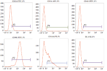
图2
流式细胞仪鉴定人根尖乳头干细胞
Figure 2
Flow cytometry identification of human stem cells from apical papilla
The expression rates of CD24, CD34, CD45, CD90, and CD146 in SCAPs were respectively 15.57%, 0.81%, 0.35%, 98.70%, 42.42%
2.2 CCK-8检测LED红光对SCAPs增殖的影响
根据CCK-8检测的结果绘制每组hSCAPs的生长曲线,见图3a。与对照组相比,在光照后第1、3、5、7和9天,实验组均显著促进hSCAPs的增殖。光照后第1天(1 J/cm2组vs.对照组,P=0.002;3 J/cm2组vs.对照组,P=0.001;5、7 J/cm2组vs.对照组,P<0.001)、第7天(F=51.110,P<0.001)、第9天(F=83.743,P<0.001),各光照组的细胞增殖能力均强于对照组。光照后第3天,3、5、7 J/cm2组的细胞增殖率明显高于对照组(F=106.942,P<0.001)。光照后第5天,仅5 J/cm2组显示出更强的细胞增殖能力(F=18.199,P<0.001)。此外,在各检测时间点的光照组之间,hSCAPs的细胞增殖率也有差异,见图3b。
图3
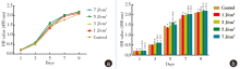
图3
LED红光对人根尖乳头干细胞增殖的影响
Figure 3
The effect of red LED on the proliferation of human stem cells from apical papilla
a: the CCK-8 growth curve of SCAPs; b: statistical differences between groups, α: vs. control group, P<0.05; β: vs. 1 J/cm2 group, P<0.05; γ: vs. 3 J/cm2 group, P<0.05; δ: vs. 7 J/cm2 group, P<0.05
2.3 ALP染色和ALP定量检测LED红光对hSCAPs成骨分化的影响
LED红光照射后第7天和第14天,ALP染色结果显示光照组较对照组着色较明显,且5 J/cm2组着色最深;同时,光照后第14天各组较第7天染色深,见图4a。ALP活性检测结果显示在光照后第7天和第14天,光照组ALP活性高于对照组;光照后第7天,3、5和7 J/cm2组促进hSCAPs的ALP活性(F=9.380,3 J/cm2组vs.对照组,P=0.006;5 J/cm2组vs.对照组,P<0.001;7 J/cm2组vs.对照组,P=0.008);在各光照组之间,5 J/cm2组ALP活性最高(5 J/cm2组vs. 1 J/cm2组,P=0.002;5 J/cm2组vs. 3 J/cm2组,P=0.043;5 J/cm2组vs. 7 J/cm2组,P=0.032);光照后第14天,仅3 J/cm2和5 J/cm2组促进hSCAPs的ALP活性(F=36.867,3 J/cm2组vs.对照组,P=0.035;5 J/cm2组vs.对照组,P<0.001),5 J/cm2组较其他光照组显著提高ALP活性(P<0.001);同时,光照后第14天各组都显示出比第7天更高的ALP水平,见图4b。
图4
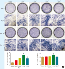
图4
LED红光对人根尖乳头干细胞成骨分化早期的影响
Figure 4
The effect of red LED on the early stage of osteogenic differentiation of human stem cells from apical papilla
a: macroscopic images and microscopic images (× 40) after ALP staining on 7 days and 14 days; b: ALP activity of SCAPs after irradiation on days 7 and 14. *: vs. control group, P<0.05; #: vs. the other irradiation groups, P<0.05
2.4 茜素红定量检测LED红光对hSCAPs矿化的影响
图5
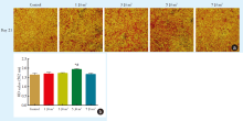
图5
LED红光对人根尖乳头干细胞成骨分化晚期的影响
Figure 5
Effect of red LED on the late stage of osteogenic differentiation of human stem cells from apical papilla
a: microscopic images (× 40) after alizarin red staining; b: the result of quantitative detection of alizarin red. * : vs. control group, P<0.05; #: vs. the other irradiation groups, P<0.05
2.5 RT-PCR检测LED红光对hSCAPs成骨相关基因表达水平的影响
在光照后第7天,5 J/cm2组较对照组上调hSCAPs中成骨相关基因ALP(F=784.697,P<0.001),OCN(F=3 563.798,P<0.001),OPN(F=511.775,P<0.001),BSP(F=150.117,P<0.001)和Runx2(F=713.872,P<0.001)的表达,差异有统计学意义。光照后第14天,5 J/cm2组上调hSCAPs中成骨相关基因ALP(F=601.905,P<0.001)、OCN(F=779.152,P<0.001)、OPN(F=1120.033,P<0.001)、BSP(F=1736.171,P<0.001)和Runx2(F=206.170,P<0.001)的表达,差异有统计学意义,见图6。
图6
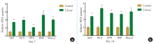
图6
LED红光对人根尖乳头干细胞成骨相关基因表达的影响
Figure 6
Effect of red LED on the expression of osteogenic related genes of human stem cells from apical papilla
a: the expression of osteogenic related genes of hSCAPs on day 7; b: the expression of osteogenic related genes of hSCAPs on day 14. ALP: alkaline phosphatase; OCN: osteocalcin; OPN: osteopontin; BSP: bone sialoprotein; Runx2: Runt-related transcription factor 2; *: vs. control group, P<0.05
2.6 Western blot检测LED红光对hSCAPs成骨相关蛋白表达水平的影响
Western blot结果显示,5 J/cm2组在第7天ALP(F=172.997,P<0.001)、OCN(F=465.951,P<0.001)、OPN(F=207.232,P<0.001)、BSP(F=209.934,P<0.001)和Runx2(F=585.258,P<0.001)蛋白的表达上调,差异具有统计学意义。5 J/cm2组在第14天上调ALP(F=110.244,P<0.001)、OCN(F=182.052,P<0.001)、OPN(F=38.148,P=0.003)、BSP(F=65.461,P=0.001)和Runx2(F=79.046,P=0.001)蛋白的表达,差异具有统计学意义,见图7。
图7
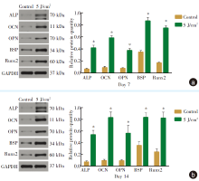
图7
LED红光对人根尖乳头干细胞成骨相关蛋白表达的影响
Figure 7
Effect of red LED on the expression of osteogenic related proteins of human stem cells from apical papilla
a: the result of electrophoretic bands and expression of osteogenic related proteins of hSCAPs on day 7; b: the result of electrophoretic bands and expression of osteogenic related proteins of hSCAPs on day 14. ALP: alkaline phosphatase; OCN: osteocalcin; OPN: osteopontin; BSP: bone sialoprotein; Runx2: Runt-related transcription factor 2; *: vs. control group, P<0.05
3 讨论
LED红光已被证明具有多种生物调节作用,如减轻炎症和促进伤口愈合[17],具有价格低廉、使用寿命长、电路实现简单、应用安全等优点,仪器便于携带,可以作为在家中进行光动力治疗的工具[6],可以通过多种方式影响不同干细胞的增殖和成骨分化[18,19]。现阶段LED红光对牙源性干细胞增殖和成骨分化的影响逐渐引起关注,但其对hSCAPs生物学特性的影响研究较少。Horvát-Karajz等[20]发现细胞经光照后的光生物学效应可以持续48 h。Marques等[21]发现1.2~7.5 J/cm2的红光能对干细胞的细胞存活率和增殖产生积极效应。同时本课题组前期实验发现1、3、5 J/cm2的LED红光对牙源性间充质干细胞有积极作用[10]。故本实验选择1、3、5、7 J/cm2 LED红光,每48 h对细胞进行一次照射,探讨LED红光对hSCAPs增殖及成骨分化的影响,以期为光生物疗法在牙组织工程中的应用筛选适宜的光学参数,为hSCAPs作为牙再生种子细胞提供实验数据。
Hamblin等[6]认为光生物调节作用存在双相剂量反应,即低剂量光照具有促进作用,高剂量光照产生抑制作用,可能是细胞色素C氧化酶光受体吸收LED红光后被激活,提供更高速率的氢质子泵入线粒体,产生能量,并催化产生活性氧(reactive oxygen species,ROS)促进细胞增殖。Ferreira等[9]报道5 J/cm2红光促进牙髓干细胞增殖,Marques等[21]发现5 J/cm2红光照射乳牙牙髓干细胞的存活率和增殖率较高,Yamauchi等[22]发现8 J/cm2对PDLSCs增殖促进效果最显著。本实验中,增殖实验结果也显示LED红光在hSCAPs增殖早中晚期均有不同程度的促进作用,5 J/cm2红光照射对hSCAPs的促进作用最显著,提示LED红光照射对不同干细胞产生的光生物学效应有微小差异。
本实验发现在成骨诱导条件下,LED红光促进hSCAPs的成骨分化及钙结节的形成,其中5 J/cm2组在第7、14和21天均有良好的促进作用,表明5 J/cm2 LED红光可以在hSCAPs早中晚期成骨中均发挥有利作用。根据以上结果,笔者选择5 J/cm2组与对照组进行PCR和western blot实验探讨相关机制。ALP和Runx2是成骨分化早期的标志[23,24],OCN在成骨细胞分化晚期表达[25]。OPN的表达与骨形成/吸收相关,是成骨分化中晚期的标志[26]。BSP在骨形成开始时、成骨分化后期和矿化早期表达[27]。结果显示5 J/cm2 LED红光能上调ALP、Runx2、OCN、OPN、BSP基因和蛋白的表达,进一步提示LED红光促进SCAPs成骨分化。Li等[28]报道LED红光促进大鼠骨髓间充质干细胞成骨分化,Ruan等[18]发现LED红光具有促进人骨髓间充质干细胞成骨分化的作用,与本研究结果一致。但Pagin等[29]提出LED红光不影响前成骨细胞MC3T3细胞的分化,这可能与LED红光照射曝光量等光学参数不一致有关,也可能与LED红光选择性促进干细胞成骨分化有关。
综上所述,本实验探讨LED红光对hSCAPs增殖和成骨分化的量效关系,发现在模拟成骨诱导环境中,LED红光促进hSCAPs体外增殖和成骨分化,其中5 J/cm2的光照效果最显著,为促进hSCAPs和光生物调节疗法在组织工程和干细胞治疗中的应用提供了新的研究基础。
【Author contributions】 Su YT performed the experiments and wrote the article. Hou L, Jiang B, Zheng GZ, Liu Y and Wang Y performed the experiments and revised the article. All authors read and approved the final manuscript as submitted.
官网
参考文献
Dental tissue-derived human mesenchymal stem cells and their potential in therapeutic application
[J].
Mesenchymal stem cell-mediated functional tooth regeneration in swine
[J].
牙源性干细胞储存和临床应用的研究进展
[J].
Progress in storage and clinical application of dental stem cells
[J].
Proliferation, odontogenic/osteogenic differentiation, and cytokine production by human stem cells of the apical papilla induced by biomaterials: a comparative study
[J].
Stem cells from the apical papilla: a promising source for stem cell-based therapy
[J].
Mechanisms and applications of the anti-inflammatory effects of photobiomodulation
[J].
Comparison and evaluation of the low-level laser and the red and blue LED effects on wound healing in rabbit
[J].
Effect of photobiomodulation therapy on the increase of viability and proliferation of human mesenchymal stem cells
[J].
We have investigated how low intensity laser irradiation emitted by a multiwave-locked system (MLS M1) affects the viability and proliferation of human bone marrow mesenchymal stem cells (MSCs) depending on the parameters of the irradiation.Cells isolated surgically from the femoral bone during surgery were identified by flow cytometry and cell differentiation assays. For irradiation, two wavelengths (808 and 905 nm) with the following parameters were used: power density 195, 230, and 318 mW/cm, doses of energy 3, 10, and 20 J (energy density 0.93-6.27 J/cm ), and in continuous (CW) or pulsed emission (PE) (frequencies 1,000 and 2,000 Hz).There were statistically significant increases of cell viability and proliferation after irradiation at 3 J (CW; 1,000 Hz), 10 J (1,000 Hz), and 20 J (2,000 Hz).Irradiation with the MLS M1 system can be used in vitro to modulate MSCs in preparation for therapeutic applications. This will assist in designing further studies to optimize the radiation parameters and elucidate the molecular mechanisms of action of the radiation. Lasers Surg. Med. © 2019 Wiley Periodicals, Inc.© 2019 Wiley Periodicals, Inc.
Short-term evaluation of photobiomodulation therapy on the proliferation and undifferentiated status of dental pulp stem cells
[J].
Irradiation with red light-emitting diode enhances proliferation and osteogenic differentiation of periodontal ligament stem cells
[J].
Recent trends in multipotent human mesenchymal stem/stromal cells: learning from history and advancing clinical applications
[J].
Local injection of allogeneic stem cells from apical papilla enhanced periodontal tissue regeneration in minipig model of periodontitis
[J].
Impact of remnant healthy pulp and apical tissue on outcomes after simulated regenerative endodontic procedure in rat molars
[J].When regenerative endodontic procedures (REPs) are performed on immature teeth diagnosed with pulp necrosis and apical periodontitis, various healing patterns occur. Furthermore, infected immature teeth with endodontic disorders often exhibit some remnant pulp and apical tissue. Therefore, this study investigated the impact of remnant healthy or fully functional pulp and apical tissue on healing patterns after REPs. Simulated REPs were performed on non-infected immature rat molars with different amounts of remnant pulp and apical tissue. Healing patterns in these teeth were assessed after 28 days. Teeth with 0.81-0.91 mm of remnant pulp healed with pulp-like tissue, dentin, and osteodentin-like dentin-associated mineralized tissue (OSD-DAMT); teeth with 0.60-0.63 mm of remnant pulp healed with pulp-like tissue and OSD-DAMT; teeth with 0.13-0.43 mm of remnant pulp healed with periodontal ligament (PDL)-like tissue, OSD-DAMT, and cementum-like dentin-associated mineralized tissue (CEM-DAMT); and teeth with disorganization of pulp and apical tissues at 0.15-0.38 mm beyond the root apex healed with PDL-like tissue, CEM-DAMT, and intracanal bone (IB). Loss of Hertwig's epithelial root sheath was observed with IB formation. These results showed that four distinct healing patterns occurred after REPs, depending on the preoperative amount of remnant healthy pulp and apical tissue.
Exosomes derived from stem cells from the apical papilla promote dentine-pulp complex regeneration by inducing specific dentinogenesis
[J].
SFRP2 promotes stem cells from apical papilla-mediated periodontal tissue regeneration in miniature pig
[J].
Evaluation of odonto/osteogenic differentiation potential from different regions derived dental tissue stem cells and effect of 17β-estradiol on efficiency
[J].
Photobiomodulation therapy reduces acute pain and inflammation in mice
[J].
Irradiation by high-intensity red light-emitting diode enhances human bone marrow mesenchymal stem cells osteogenic differentiation and mineralization through Wnt/β-catenin signaling pathway
[J].
The effect of LED photobiomodulation on the proliferation and osteoblastic differentiation of periodontal ligament stem cells: in vitro
[J].The aim of this study was to investigate the influence of three different light-emitting diode (LED) wavelengths on the proliferation and osteoblastic differentiation of periodontal ligament stem cells (PDLSCs) in vitro.PDLSCs seeded on 96- and 24-well plates, for proliferation and osteoblastic differentiation, respectively, were irradiated daily by LED light with peak emission wavelengths of 630, 680, and 830 nm at constant energy densities of 3.5 J/cm. Cultures were grown for 8 days for the proliferation assay, 10 days for the alkaline phosphatase (ALP) assay, and 28 days for Alizarin red staining. Mitochondrial activity, ALP enzyme level, and the ability to form calcium phosphate deposits were measured and compared across cultures.Results obtained from statistical analysis of the experimental data indicated that the rate of proliferation (P < 0.05) in 830-nm irradiated cultures were significantly higher than the control samples at day 6 and 8; whereas, for the 630- and 680-nm groups, test results showed lower proliferation rates at day 8. For osteoblastic differentiation, significantly greater mineralization than the control samples was detected in the red-light groups (630 and 680 nm) during the late differentiation period (P < 0.001), which was supported by a higher ALP activity of the 630- and 680-nm groups in the early stage (P < 0.01).The results of this study demonstrate that the PDLSCs responded differently to specific LED wavelengths. For enhancing cellular proliferation, 830-nm LED irradiation was more effective. On the other hand, the wavelengths of 630 and 680 nm were better for stimulating osteoblastic differentiation.Copyright © 2021 World Federation of Orthodontists. Published by Elsevier Inc. All rights reserved.
In vitro effect of carboplatin, cytarabine, paclitaxel, vincristine, and low-power laser irradiation on murine mesenchymal stem cells
[J].Mesenchymal stem cells (MSCs) are promising for use in regenerative medicine. Cytostatics can decrease, but low-power laser irradiation (LPLI) can increase the growth of MSCs. The interaction of LPLI, MSCs and cytostatics is not known. This study investigated the effect of four cytostatics (carboplatin, cytarabine, paclitaxel, vincristine), LPLI, and combination of a cytostatic drug and LPLI on murine MSCs (mMSCs).MMSCs were exposed to LPLI (660 nm diode laser; 60 mW output power; range of power density: 76-156 mW/cm(2); range of energy density: 1.9-11.7 J/cm(2)) and/or a cytostatic drug (carboplatin: 2, 10, 50; cytarabine: 0.4, 10, 50; paclitaxel: 0.4, 2, 10; vincristine: 0.02, 0.1, 0.5 microg/ml, respectively). Cell proliferation was measured after 24, 48, or 72 hours incubation.LPLI at 1.9 J/cm(2) dose increased the proliferation rate with 41% after 48 hours. However, 11.7 J/cm(2) LPLI caused 42% inhibition and cytostasis was still detectable after 72 hours. LPLI caused equivalent stimulation in single or in divided doses (3.8 vs. double 1.9 J/cm(2) in a 24-hour period). The cytotoxicity of 50 microg/ml carboplatin was eliminated, the inhibitory power of 0.1 microg/ml vincristine was attenuated by 1.9 J/cm(2) LPLI even 3 days post-treatment (attenuation >10%). The 11.7 J/cm(2) LPLI enhanced the cytotoxicity of 50 microg/ml cytarabine (from 48% to 73%) and 10 microg/ml paclitaxel (from 37% to 78%). Combination of the ineffective 0.4 microg/ml cytarabine or paclitaxel with the inhibitory 11.7 J/cm(2) LPLI exhibited stronger inhibition than the 11.7 J/cm(2) LPLI alone (69% and 69% vs. 42%).Low energy density of LPLI increases and high energy density of LPLI decreases the proliferation of mMSCs. Furthermore, LPLI can prevent or attenuate some drug's cytotoxicity and amplify others'. The result depends on the applied energy density, on the type and concentration of the cytostatics.Copyright 2009 Wiley-Liss, Inc.
A preliminary comparison between the effects of red and infrared laser irradiation on viability and proliferation of SHED
[J].
High-power, red-light-emitting diode irradiation enhances proliferation, osteogenic differentiation, and mineralization of human periodontal ligament stem cells via ERK signaling pathway
[J].
Alkaline phosphatase: structure, expression and its function in bone mineralization
[J].
Regulation of proliferation, differentiation and functions of osteoblasts by Runx2
[J].
Functions of osteocalcin in bone, pancreas, testis, and muscle
[J].
Osteopontin in bone metabolism and bone diseases
[J].
Osteogenic differentiation and immune response of human bone-marrow-derived mesenchymal stem cells on injectable calcium-silicate-based bone grafts
[J].
Red-light light-emitting diode irradiation increases the proliferation and osteogenic differentiation of rat bone marrow mesenchymal stem cells
[J].


